Image courtesy of renjith krishnan at FreeDigitalPhotos.net

The Respiratory System
The respiratory system is responsible for supplying the cells with the oxygen required for aerobic cellular respiration, and removing the waste product CO₂
Oxygen is contained in the air and it enters the lungs by a process called ventilation (breathing). Other functions of the respiratory system include vocalization (speech) and olfaction (smell)
The main organs of the respiratory system are the:
-
Nasal cavity
-
Pharynx
-
Larynx
-
Trachea
-
Bronchi
-
Lungs (bronchioles and alveoli)
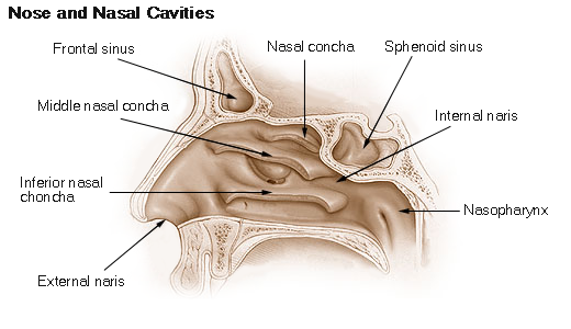
The Nasal Cavities
The nasal cavity is a hollow space within the nose and skull that is lined with hairs and mucous membrane. Its function is to warm, moisturize, and filter air entering the body before it reaches the lungs.
Hairs and mucus lining the nasal cavity help to trap dust, mould, pollen etc. before they reach the lungs. The sensory organs of smell are also found in the roof of the nasal cavity.
The Pharynx
The pharynx or throat is the passageway leading from the mouth and nose to the oesophagus and larynx. It permits the passage of swallowed solids and liquids into the oesophagus, or gullet, and conducts air to and from the trachea (windpipe) during inspiration and expiration (breathing).
The Larynx
At the top of the trachea is your larynx (voice box), that joins the pharynx with the trachea. It consists of several cartilage tissues and lies just below the hyoid bone. It contains the vocal cords which allow the body to produce the sounds of speech and singing. At the top of the larynx, there is a small flap called the epiglottis and its function is to close the entrance to the larynx to stop you chocking on your food.
The trachea and bronchi
The trachea (windpipe) is a cylindrical tube which is about ¾ inch in diameter and 4-5 inches long and runs in front of the oesophagus. It is a flexible tube that extends from the cricoid cartilage of the larynx into the thorax, where it divides into two smaller tubes called the left and right bronchi.
It is lined with ciliated columnar epithelial cells that secrete mucous to trap dust particles and is surrounded by C shaped rings made of hyaline cartilage that provide support and protection. The rings don’t go all the way around because it would hamper the passage of food due to the oesophagus stretching as the bolus of food passes down.
At the end of the trachea, the airway splits into left and right branches and are known as the primary bronchi or left and right main bronchus. The left and right bronchus run into each lung before branching off into smaller secondary bronchi, each supplying the different lobes of the lungs.
The main function of the trachea and bronchi is to act as channels for air to reach the lungs, but it also helps condition the inspired air.
The Lungs
The human body has two lungs that start slightly above the clavicles and rest on the diaphragm. Each lung has a hilum (root) where the main bronchus, main blood and lymphatic vessels and nerves enter and leave the lungs.
The lungs are divided into lobes (the right is bigger with three lobes, whilst the left has only two lobes because of the position of the heart). They include a network of branching tubes called bronchioles. At the end of the bronchioles are the alveoli, where gaseous exchange takes place.
There are about 300 million alveoli in the lungs. They are very thin (one cell thick), which speeds up diffusion of oxygen and carbon dioxide, as there is less distance to travel. The alveoli are surrounded by blood capillaries, and this is where gaseous exchange occurs.
Pleural Membranes
There is a membrane that attaches to each lung, and another that lines the inner wall of the chest. Each membrane consists of simple squamous epithelial cells that secrete a slippery serous fluid into space between them. This allows the membranes to slide on each other, reducing friction. They also can’t be pulled apart from each other, so when the chest wall moves during breathing, the lungs move with it.
Ventilation
Ventilation is the mechanical process of breathing. Inspiration is the process of drawing air into the lungs and expiration is the removal of air from the lungs.
During inspiration the intercostal muscles contract, causing the ribs to move upwards and outwards resulting in the chest getting bigger from side to side.
Image by Anatomography - en:Anatomography (setting page of this image), CC BY-SA 2.1 jp, https://commons.wikimedia.org/w/index.php?curid=22754580
The diaphragm flattens causing the chest to get bigger from top to bottom. This increases the volume inside the chest, causing the pressure to drop. This relationship between pressure and volume is called Boyle's Law.
Due to the pressure inside the lungs now being lower than the atmospheric pressure, suction is created causing air to flow into the lungs.
Image by OpenStax College [CC BY 3.0 (http://creativecommons.org/licenses/by/3.0)], via Wikimedia Commons
During expiration the intercostal muscles relax, causing the ribs to move downwards and inwards resulting in the chest getting smaller from side to side.
The diaphragm returns to its resting dome shape causing the chest to get smaller from top to bottom. This decreases the volume inside the chest, causing the pressure to increase. Due to the pressure inside the lungs now being higher than the atmospheric pressure, the air is blown out of the lungs.
Gaseous Exchange
Gaseous exchange occurs between the alveoli of the lungs and the capillaries of the cardiovascular system. There are about 300 million sphere- shaped alveoli in each lung providing a large surface area, all surrounded by a large network of capillaries.
The structure of the alveoli and capillaries make them ideal for diffusion. Both structures are very thin (simple squamous epithelial cells), therefore the distance for gases to move between membranes is very small.
Image by Patrick J. Lynch, medical illustrator (Patrick J. Lynch, medical illustrator) [CC BY 2.5 (http://creativecommons.org/licenses/by/2.5)], via Wikimedia Commons
Respiratory surfaces have to remain moist to facilitate the diffusion of O₂, hence the alveoli have a fluid lining. The presence of surfactant in the fluid lining also prevents the alveoli from collapsing.
Image by National Heart Lung and Blood Institute (National Heart Lung and Blood Institute) [Public domain], via Wikimedia Commons
Gaseous exchange occurs by a process called diffusion; that is the movement of molecules from an area of high concentration to an area of low concentration.
At a cellular level, there will be a spontaneous movement of oxygen and carbon dioxide in and out of the cell. Oxygen and carbon dioxide are small molecules so they can easily pass through the cell membrane.
When oxygen flows into a cell it will always be used up in cellular respiration, therefore the concentration inside will always be low meaning it will continue to diffuse into the cell.
Carbon dioxide is a waste product of cellular respiration so the concentration will always be higher inside the cell. Carbon dioxide will therefore always diffuse out of the cell.
When we breathe in there will be a higher concentration of oxygen in the alveoli, than their surrounding capillaries. The oxygen will, therefore, move by the process of diffusion from where it is high in concentration (alveoli) into the capillaries where it is low in concentration.
At the same time, the carbon dioxide that is higher in concentration in the capillaries will move into the alveoli, where it is lower in concentration, to be breathed out.